
What gets “frozen”?
Where do the “adhesions” form?
What causes it?
So many questions without answers. Frozen is a bit of a misnomer – it is an inflammatory process. Attribute the term to Codman and his 1934 textbook, “The shoulder”. No adhesions form – rather, the capsule becomes thickened and conracted. The condition is usually idiopathic, which means that we aren’t smart enough to know exactly why it happens.
Etiology – “Idiopathic” frozen shoulder
The condition usually occurs without a known inciting event, although many patients will recall when they first felt pain. Often, patients will say the problem began when they reached into the back seat of the car. It is likely the process had already begun when they reached and felt the discomfort.
Typical age presentation is between 40 to 60 years old, with a slightly elevated risk in the female population.1 FS is a surprisingly common condition, affecting between 5 and 20 percent of the population at some point in their lives.
There is a definitive link to endocrine conditions. If I suspect that someone has a frozen shoulder, the first questions that I ask are often “do you have diabetes” and “do you have thyroid problems”? We don’t know why. We do know that if these conditions are present, treatment is a bit more challenging and prolonged. Higher blood sugar levels, and elevated HbA1c levels also make FS harder to treat.2
Other associated elements of the history include a history of recent neck surgery, breast surgery, or a history of a frozen shoulder on the other side.
Etiology – other
Sometimes prior injury or trauma, such as a fracture around the shoulder, will lead to a stiff and painful shoulder. This is considered a “post-traumatic” stiff shoulder – treatment and outcomes of care are quite different. Consideration of this particular type of FS will occur in a different post.
Shoulder arthritis will also lead to stiffness and loss of mobility. Although patients with stiffness and arthritis may benefit from a similar treatment algorithm as those with FS, expectations for response are vastly different. Naming “frozen shoulder” as the diagnosis in patients with arthritis is wrong, and should be avoided.
Pathology
Residents in training learn that the process is called “fibroblastic proliferation.” It means that cells in the capsule that make collagen become a bit too active. They produce abundant amounts of this material (especially type III collagen3), and it leads to a thickening of the capsule. A definite inflammatory component is involved in the earlier phases of the disease. The capsular tissue has a typical inflamed appearance. We are beginning to understand which cytokines, growth factors, and inflammatory mediators are involved in the process (TGF-B, IL-6, MMP, TNF-alpha, PDGF, among others). Hence, pain is one of the most common presenting complaints. Inflammation hurts. It wakes people at night. And attempts to stretch the shoulder will be stretching this inflammatory tissue.

Stages
Three stages of the disease are classically described: freezing, frozen, and thawing. However, these stages are not distinct periods of time with different pathological and histological findings. Additionally, the length of each phase can be quite variable. We tell patients that spontaneous resolution is possible, but it can take 2 years or longer for this to happen without intervention.
The freezing stage presents with pain, but relative preservation of motion. Night pain, gradually worsening symptoms, and some confusion about the diagnosis can occur. Physical therapy referrals can be made. Often, if the shoulder has not yet lost mobility, typical “rotator cuff” exercises are prescribed. Unfortunately, this set of exercises is not beneficial for patients with FS. In fact, rotator cuff exercise can exacerbate the problem. As such, patients in this stage will commonly say that “physical therapy didn’t help”.
The frozen stage is when the fibrosis of the capsule has occurred. A simple history and physical exam will quickly confirm the diagnosis. Xrays are normal. MRI findings are variable – but often show age related changes of the rotator cuff and labrum. This only adds confusion to the diagnosis. MRI should largely not be part of the typical workup for these patients, due to the high prevalence of “abnormal” findings4. One characteristic MRI finding is loss of the normally lax and patulous inferior recess on the coronal images. But this is not useful from a diagnostic standpoint.
The thawing stage is recognized by the restoration of mobility and the diminution of pain. The inflammatory component has resolved, and the capsule returns to its normal lax state.
Treatment – conservative
Fortunately, FS is quite responsive to treatment. I often talk about the 2 main components in my discussion with patients: the inflammatory component and the capsular fibrosis component.
The inflammatory component is highly responsive to an intra-articular corticosteroid injection. This medication effectively modulates the inflammatory process, and I see about 80% of patients achieve 80% relief of pain within a month. If only 1 intervention could be chosen, this might be the most useful to restore comfort and function5. It seems the key is injecting into the joint space, which can be challenging when done without imaging guidance. Sethi showed in 2005 that only 25% of attempted injections from an anterior approach actually made it into the joint6. The same author showed that the anterior approach was more effective than posterior (80% vs 50%) without guidance7. Further evidence that anterior injection is the most predictable was shown in 2010 by Tobola8. 65% were successful vs 46% from posterior. These studies were done in patients without frozen shoulder, which is a more challenging scenario to successful enter the glenohumeral joint. As such, I typically arrange for guided injection – either ultrasound or fluoroscopic (xray) guidance. Ultrasound can produce 97% accuracy9. Fluoroscopy should produce 100% accuracy, but comes with some inconvenience and added cost for the patient.
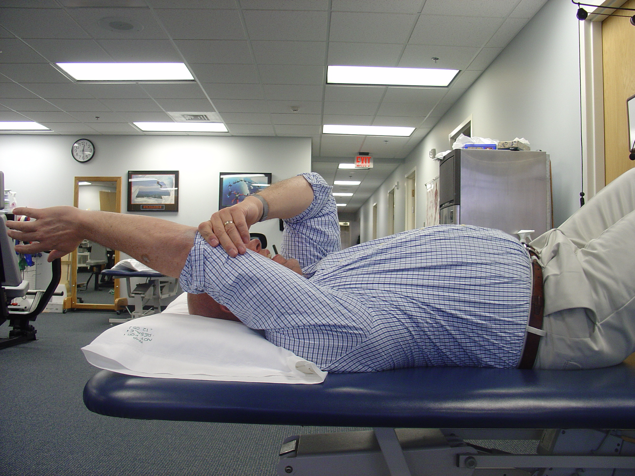
The stiff capsule is responsive to a program of frequent stretching throughout the day10. Recommendation is for 5 session of several minutes each, holding each stretch for at least 30-45 seconds. “Frequency, not forcefulness” was taught by my mentors at the University of Washington. A program of “4-quadrant” stretching can bring 80% improvement in 80% of patients within a month, when combined with intra-articular injection.


Treatment – surgical
For those patients who do not respond to the above interventions. 2 surgical options are available. I have become accustomed to following the algorithm learned at the University of Washington, based on experience of the surgeons at that institution.
“Manipulation under anesthesia”, or MUA, involves a stretch of the shoulder while the patient is under anesthesia in the operating room. MUA is a very quick procedure that allows stretching of the different parts of the capsule by pushing the shoulder into its extremes in all directions (roughly following the “4 quadrant” pattern). This is typically followed by formal work with physical therapy for a few weeks after to prevent recurrence. This procedure works well for those truly idopathic cases that are not associated with endocrine problems, prior surgery, trauma, etc. If any of those associations are present, I opt instead for arthroscopic release.
Arthroscopic release involves a relative short and easy procedure where the capsule is released with small tools under direct visualization with use of a small camera that is placed into the shoulder. Recovery is often fairly uncomplicated. Immobilation is not utilized. Physical therapy is prescribed for a few weeks.

The biggest risk of surgical care is recurrence of the frozen shoulder, although it is relatively uncommon. There seems to be some increased risk of recurrence if performed too early in the disease course. Hence, we always start with conservative treatments, and typically wait 6-9 months prior to consideration of surgery.
Additionally, development of frozen shoulder on the other side can occur (up to 50% of the time). Thus, successful care of frozen shoulder on one side can lead to development of FS on the other shoulder.
Summary
Frozen Shoulder is a common condition, with an unknown cause. The vast majority of patients will respond quite readily to a single intra-articular injection (best done under guidance) combined with a simple “4 quadrant” stretching program done frequently throughout the day. For those patients that do no respond, an MUA or arthroscopic release will bring return of normal comfort and function with low risk of recurrence or complication.
References
- Kingston K, Curry EJ, Galvin JW, Li X. Shoulder adhesive capsulitis: epidemiology and predictors of surgery. J Shoulder Elbow Surg. (2018) 27:1437–43. doi: 10.1016/j.jse.2018.04.004 ↩︎
- Chan JH, Ho BS, Alvi HM, Saltzman MD, Marra G. The relationship between the incidence of adhesive capsulitis and hemoglobin A1c. J Shoulder Elbow Surg. (2017) 26:1834–7. doi: 10.1016/j.jse.2017.03.015 ↩︎
- Rodeo SA, Hannafin JA, Tom J, Warren RF, Wickiewicz TL. Immunolocalization of cytokines and their receptors in adhesive capsulitis of the shoulder. J Othop Res. 1997;15:427–436. doi: 10.1002/jor.1100150316. ↩︎
- Dimitriou D, Winkler E, Zindel C, Grubhofer F, Wieser K, Bouaicha S. Is routine magnetic resonance imaging necessary in patients with clinically diagnosed frozen shoulder? Utility of magnetic resonance imaging in frozen shoulder. JSES Int. 2022 Jun 11;6(5):855-858. doi: 10.1016/j.jseint.2022.05.009. PMID: 36081696; PMCID: PMC9446195. ↩︎
- Challoumas D, Biddle M, McLean M, Millar NL. Comparison of Treatments for Frozen Shoulder: A Systematic Review and Meta-analysis. JAMA Netw Open. 2020 Dec 1;3(12):e2029581. doi: 10.1001/jamanetworkopen.2020.29581. PMID: 33326025; PMCID: PMC7745103. ↩︎
- Sethi PM, Kingston S, Elattrache N. Accuracy of anterior intra-articular injection of the glenohumeral joint. Arthroscopy. 2005 Jan;21(1):77-80. doi: 10.1016/j.arthro.2004.09.009. PMID: 15650670. ↩︎
- Sethi PM, El Attrache N. Accuracy of intra-articular injection of the glenohumeral joint: a cadaveric study. Orthopedics. 2006 Feb;29(2):149-52. doi: 10.3928/01477447-20060201-01. PMID: 16485459. ↩︎
- Tobola A, Cook C, Cassas KJ, Hawkins RJ, Wienke JR, Tolan S, Kissenberth MJ. Accuracy of glenohumeral joint injections: comparing approach and experience of provider. J Shoulder Elbow Surg. 2011 Oct;20(7):1147-54. doi: 10.1016/j.jse.2010.12.021. Epub 2011 Apr 13. PMID: 21493103. ↩︎
- Kuratani K, Tanaka M, Hanai H, Hayashida K. Accuracy of shoulder joint injections with ultrasound guidance: Confirmed by magnetic resonance arthrography. World J Orthop. 2022 Mar 18;13(3):259-266. doi: 10.5312/wjo.v13.i3.259. PMID: 35317253; PMCID: PMC8935327. ↩︎
- Griggs SM, Ahn A, Green A. Idiopathic adhesive capsulitis. A prospective functional outcome study of nonoperative treatment. J Bone Joint Surg Am. 2000 Oct;82(10):1398-407. PMID: 11057467. ↩︎
Leave a Reply